The role of glial cells in Alzheimer's and potential treatments
Yathushaa Navinan • 2024-06-28
Alzheimer’s is a neurodegenerative disease commonly known for causing memory loss but can also cause behavioral changes and cognitive deterioration
Alzheimer’s is a neurodegenerative disease commonly known for causing memory loss but can also cause behavioral changes and cognitive deterioration (1). It is the most common cause of dementia in the UK, with dementia referring to “a group of symptoms associated with an ongoing decline of brain functioning”, affecting mental abilities such as memory, thinking skills and others (2).
Current research on the development and treatment of Alzheimer’s disease is overwhelmingly focused on the role of beta-amyloid protein. The amyloid cascade hypothesis has been the leading explanation for the development of Alzheimer’s for decades, stating that difficulty in processing the beta-amyloid protein causes production of plaques in the brain that is toxic to neurons and synapses and therefore causes cell death, leading to dementia (3). This has caused most research to focus on developing treatments based on this hypothesis, a high-profile recent example being the development of the new antibody drug donanemab (1). However, in recent years, the amyloid cascade hypothesis has been questioned, causing many researchers to propose various hypotheses such as the pathogenic hypothesis, the vascular hypothesis and the declined brain metabolic activity independent of etiology hypothesis. This article will focus on a few hypotheses put forward concerning the role of glial cells in the development of Alzheimer’s.
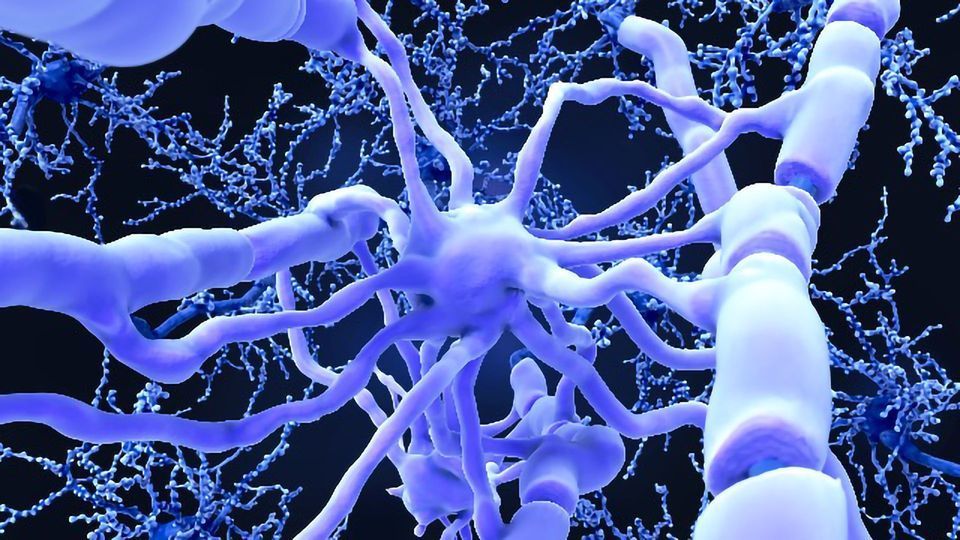
The brain consists of two types of cells: neuronal and glial cells. The function of glial cells includes the maintenance of homeostasis within the neuronal vicinity (concentration of ions, neurotransmitters etc.) and aid in the formation of myelin sheaths around neuron axons, which are responsible for cell-to-cell signaling and maintaining the function of synapses (4). Microglia, oligodendrocytes, polydendrocytes, astrocytes, Schwann cells, satellite cells, ependymal cells and enteric cells are among the many different types of glial cells (4). Of these, microglia and astrocytes have the most focus in glial cell research for Alzheimer’s disease, but some research has also been dedicated to the role of oligodendrocytes and polydendrocytes as well
Credit: Simple psychology - https://www.simplypsychology.org/glial-cells.html
Microglia
Microglia plays an important role in the neuroprotection of nerve cells and tissue cell mechanisms in response to neurodegenerative diseases. For example, in Alzheimer’s, beta-amyloid proteins aggregate glial cells such as microglia, which remove beta-amyloid proteins and reduce accumulation of these proteins that lead to plaque formation (5). However, one study stated that the “phagocytic function of microglia is activated in the brain in response to brain damage and diseases” (5). This means microglial cells are releasing neurotoxins which result in neuronal damage and thus symptoms of Alzheimer’s disease. Furthermore, clinical trials have found no evidence of microglia being involved in beta-amyloid protein accumulation and therefore questions the amyloid cascade hypothesis (5). However, there is evidence that microglia are responsible for the degradation of beta-amyloid proteins, causing soluble beta amyloid molecules to be more present in brain tissue compared to the cerebrospinal fluid (5). This approach suggests new potential treatments for Alzheimer’s.
In addition to this, the neuroprotective function of microglia is rendered less efficient because of neuroinflammation caused by beta-amyloid accumulation and debris from dead nerve cells (5). This is demonstrated in studies of old mice, where microglia lost the ability to recognize and degrade beta-amyloid proteins (5).
Furthermore, microglia have been found to be involved in neuroinflammation and Alzheimer's pathogenesis in the elderly, which leads to the activation of several receptors such as NDMA and GluR1, therefore leading to neuronal cell death due to dysfunction of neuronal synapses that these activated receptors cause (6). This has been demonstrated in studies of mice, where microglia-induced inflammation in the hippocampus was found to induce reduced hippocampal function and therefore cognitive decline (6).
Astrocytes:
Some studies have found astrocytes to be activated by beta-amyloid proteins, resulting in an increase in calcium ion concentrations within neuronal cells “that stimulates NADPH oxidase, causing neuronal death through oxidative stress” (5). Oxidative stress refers to an imbalance between biological molecules with high oxidizing power, and antioxidant agents produced by the body.
This increase in calcium ion concentrations may be due to impaired Ca2+ signaling in astrocytes in those with the disease (7). Intracellular calcium signals are used by astrocytes to maintain the homeostasis of neuronal circuits and regulate neuronal activity, which means the dysfunction of calcium ion signaling leads to neuronal hyperactivity in early stages of Alzheimer’s that may contribute to pathological progression of the disease (7). Supporting evidence comes from a study of astrocyte calcium signaling in patients with Alzheimer’s disease, with findings of decreased expression of the receptor ITPR2, an intracellular calcium release channel (7).
Astrocytes can also cause impaired glutamate buffering in patients with Alzheimer’s (7). Glutamate is an excitatory neurotransmitter, which means it encourages electrical activity in the brain. Glutamate receptor activation by glutamate is known to contribute to various neurological functions including memory, cognition, movement and sensation (7). Astrocytes are involved in glutamate uptake, clearing around 80 to 90% of glutamate in synapses, however differences in glutamate receptors on astrocytes in patients with Alzheimer’s disease causes reduced glutamate absorption which may contribute to pathogenesis of the disease (7). This reduction in glutamate absorption and transport is linked to beta-amyloid protein accumulation in patients, which itself is linked to neuronal cell death (7).
Treatments:
If in appropriate amounts, astrocytes can function as a neuroprotective cell in Alzheimer’s disease, as activated astrocytes increase uptake of beta-amyloid proteins and therefore reduces neuronal death and cognitive decline (5). However, the potential of astrocytes being used as a treatment option is limited, as excess amounts can cause them to serve a neurotoxic function instead, as outlined in the section above (5).
In one study using computed models of mice with Alzheimer’s disease, reduced levels of the astrocyte receptor Slc1a2 (also known as GLT-1 in humans) was found to exacerbate cognitive impairment (7). This gives rise to potential treatments for the disease involving the restoration of GLP-1 in patients, which could lead to improved cognitive function, restored synaptic integrity and reduced beta-amyloid plaques, as was found when Slc1a2 was restored in mice (7).
Another potential treatment involving astrocytes revolves around inhibiting the CaN/NFAT pathway in astrocytes (4). This reduces astrogliosis, which is the rapid increase of astrocytes to compensate for neuronal damage, and therefore cognitive functions (4). One study achieved this by using adeno-associated virus vectors that targeted hippocampal astrocytes in mice, stimulating expression of the peptide VIVIT that interfered with the CaN/NFAT pathway, and consequently led to reduced astrocyte activation (4). After several months, the mice showed improved synaptic and cognitive function, giving potential for astrocyte-based therapies for patients with Alzheimer’s (4).
In microglial cells, the gene TREM2 is most focused on when considering microglia-based treatments for the neurodegenerative disease (4). Studies have found TREM2 to be upregulated in microglia and become more pronounced as the disease progresses (4). This gene used to be identified as a risk gene for Alzheimer’s, although several recent studies have found TREM2 to encourage phagocytosis of beta-amyloid proteins (4). This leads to an increase in neuroprotection, slowing down progression of the disease and therefore improving cognitive function of patients (4). This suggests TREM2 becoming a target for future glial cell orientated gene therapies.
Conclusion
The purpose of this article was to encourage researchers to shift their focus from the betaamyloid hypothesis and focus on the role of glial cells when looking at causes and treatments of Alzheimer’s disease. However, it is important to note that several different intersectional factors are responsible for pathogenesis of this disease which were not covered in this article. This includes the role of glial cells other than microglia and astrocytes, such as oligodendrocytes, polydenrocytes and NG2-glia, as well as oxidative stress and polyamines, which all are either directly or indirectly linked to glial cells. Therefore, it is important for researchers to explore different hypotheses other than the well-established beta-amyloid hypothesis, as this will lead to a greater likelihood of more effective treatments being developed for patients with Alzheimer’s.
References
(1) P. (2024) ‘Recent developments in Alzheimer drugs’, Journey to Healthcare, 20 May. Available at: https://www.linkedin.com/pulse/recent-developments-alzheimer-drugsjourneytohealthcare-02ete/?trackingId=%2FXSIuDYuQWmjH%2FvSF1a%2F0A%3D%3D (2) Alzheimer’s disease (2021) NHS. Available at: https://www.nhs.uk/conditions/alzheimers-disease/ (3) Struble, R.G., Ala, T., Patrylo, P.R., Brewer, G.J. and Yan, X.X., 2010. Is brain amyloid production a cause or a result of dementia of the Alzheimer's type?. Journal of Alzheimer's Disease, 22(2), pp.393-399. Available at: https://content.iospress.com/articles/journal-of-alzheimersdisease/jad100846 (4) Nirzhor, S.S.R., Khan, R.I. and Neelotpol, S., 2018. The biology of glial cells and their complex roles in Alzheimer’s disease: new opportunities in therapy. Biomolecules, 8(3), p.93. Available at: https://www.mdpi.com/2218-273X/8/3/93 (5) Kim, Y.S., Jung, H.M. and Yoon, B.E., 2018. Exploring glia to better understand Alzheimer’s disease. Animal cells and systems, 22(4), pp.213-218. Available at: https://www.ncbi.nlm.nih.gov/pmc/articles/PMC6138241/#:~:text=The%20most%20com mon%20traits%20of,primarily%20on%20this%20cell%20type (6) Bandyopadhyay, S., 2021. Role of neuron and glia in Alzheimer’s disease and associated vascular dysfunction. Frontiers in aging neuroscience, 13, p.653334. Available at: https://www.frontiersin.org/articles/10.3389/fnagi.2021.653334/full (7) Kim, J., Yoo, I.D., Lim, J. and Moon, J.S., 2024. Pathological phenotypes of astrocytes in Alzheimer’s disease. Experimental & Molecular Medicine, pp.1-5. Available at: https://www.nature.com/articles/s12276-023-01148- 0#:~:text=Astrocytes%20play%20a%20role%20in,early%20stage%20of%20AD55
See More Posts
Copyright © 2021 Govest, Inc. All rights reserved.